News Release
New ways of looking inside living cells
From developing new imaging platforms, to asking new biological questions, and developing new disease diagnostics, UC San Diego bioengineering professor Lingyan Shi is pushing the boundaries of what's possible when we look inside living cells.
 |
| UC San Diego bioengineering professor Lingyan Shi |
By Becky Ham
March 29, 2021-- Energy from an exquisitely targeted photon can vibrate the chemical bond of molecules like a pluck on a guitar string. Instead of music, these vibrational frequencies allow University of California San Diego bioengineering professor Lingyan Shi to see specific molecules within a living cell.
The cutting-edge imaging technologies used by Shi, who leads the Laboratory of Optical Bioimaging and Spectroscopy at UC San Diego, are a mouthful: stimulated Raman scattering (SRS) microscopy, multiphoton fluorescence microscopy (MPF), and resonance Raman microscopy (RRS). However, they all use light of specific wavelengths to probe chemical vibrational bonds in biological tissues.
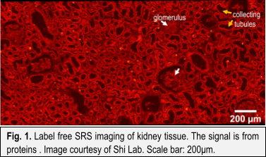 |
Different molecules such as fats, proteins, and DNA have distinct vibrational modes, and produce a subtle, yet distinct photo-spectral output. This allows researchers to identify these molecules with high chemical and spatial resolution. The powerful techniques don’t disturb living tissues, so they can be used across a variety of applications. For instance, SRS microscopy can generate a digital histological image of a biopsy in mere minutes, without the use of any chemical dyes and minimal sample preparation. Digital histology, which can mimic the staining seen in traditional histology techniques (see figure 2), may make its way into the operating room as an intraoperative technique that saves time and improves surgical performance. Another application of Raman-based technologies includes the probing of metabolic dynamics and compositions. Various tissues and even diseases like cancer are marked by distinct metabolic changes. Understanding these dynamics will aid in early detection and better understanding of such diseases and nutrition.
As one of the world’s leading experts in ultrafast optical imaging, the new Assistant Professor of Bioengineering at the UC San Diego Jacobs School of Engineering has no shortage of ideas about the potential of Raman-based technologies. “When we are developing a new technology, a new tool,” she says, “it will definitely inspire us to ask new biological questions.”
Seeing without Disturbing
 |
One of those questions is: how can we observe very small molecules in the body, without disturbing their natural functions? For her doctoral research, Shi had been tagging very small drug molecules with fluorescent probes to watch how they passed through the blood-brain barrier. But on the molecular scale, “if we consider size of the drug as a ping-pong ball, but the attached fluorescent particle was like another ping-pong ball or even bigger,” she explained. The bulky fluorescent label on the drugs was likely perturbing the drug’s behavior (such as its permeability and diffusion coefficient) in the body.
A 2014 seminar by her postdoctoral mentor Wei Min at Columbia University illustrated how stimulated Raman scattering microscopy could solve this problem. “At the time, I was very biological problem-oriented, and this was just a great tool for my research,” Shi says. “But when I got to know more and more about the technology, I realized that biological questions and imaging technology are mutually inspirational. It’s not unidirectional.”
Today, no single imaging technique reigns supreme in Shi’s lab. She and her students work with a variety of platforms, depending on the biological question at hand. Some techniques are better at seeing deep into living tissue, Shi explains, while others can probe a broader range of chemical bonds. She has also pioneered new ways of using heavy water to tag molecules with an isotope of hydrogen called deuterium (Fig. 3), to follow the metabolic activities under different physiological or pathological conditions.
As mentioned, a powerful application centers on detecting metabolic changes that occur during tumor formation, when energy-hungry cancer cells metabolize differently than healthy cells. Shi recently led a study that combined several techniques to detect early metabolic changes that distinguish triple-negative breast cancer from less aggressive cancer types.
 |
Triple-negative breast cancer predominantly affects Black and Latina women, and is difficult to detect early in the disease. “But the optical imaging technology we’re developing can tell you a lot about metabolic changes at a very early stage,” says Shi. “Early-stage diagnosis is critical to increase the survival rate among these women and reduce their cost of treatment.
“Homebuilt” Lab
Many of the imaging systems Shi and her students use are not commercially available yet, and they combine instrumentation and probes to fit their needs. “It’s like a homebuilt microscopy platform in my lab,” Shi says.
The researchers are always tinkering with the platforms to improve the image resolution and sensitivity to the optical signal. Shi holds 6 patents as a co-inventor related to optical imaging and is interested in commercializing or licensing some of these technologies.
“The application of these technologies is still in the infant stage,” she explains. “I think we need to do more educational presentations to show what kinds of information we can achieve, and to demonstrate how useful it is.”
Shi is excited to welcome research collaborators at UC San Diego, from biological scientists to genetics experts, and she is eager to mentor passionate students from underrepresented minorities in her lab, at every level from undergraduate to postdoc.
“We’re very motivated here at UCSD, and the atmosphere is great. I enjoy seeing students become aware of something they didn’t know about before,” she says. “It’s a very happy moment.”
 |
Shi joined the UC San Diego faculty in October 2019, and she says COVID-19 has made setting up a lab more difficult in some ways. At first it was challenging to source supplies, but lately she’s been more worried that the pandemic will keep her from throwing lab dinner parties and birthday celebrations that usually “bring us all together and make it feel like a family.”
“But I try to think positively about it,” Shi says. “This has also given me more time to think about the future research directions we want to explore.”
Media Contacts
Katherine Connor
Jacobs School of Engineering
858-534-8374
khconnor@ucsd.edu